
Better Science Starts with a Better Scaffold
Bio-Spun® Inserts & HTS Plates — Engineered for Results, No Reruns
Why risk wasting time, inconsistent models, and repeated experiments?
Bio-Spun® scaffolds deliver tissue-like performance that traditional cell culture tools can’t match.
Data That Pays for Itself
Higher price. Lower cost.
Even at a higher unit price, Bio-Spun® delivers lower total cost per successful experiment and superior data you can trust.

The Bio-Spun® Advantage
A scaffold proven to integrate — in vitro and in vivo.

Current Tissue Models Made with Bio-Spun® Scaffolds
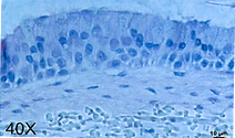
Airway Model
H&E-stained cross section of full-thickness human bronchial epithelial model produced on Bio-Spun® PET Scaffold. The scaffold is populated with pulmonary fibroblasts which produce a fully human derived subepithelial matrix component. The fully developed pseudostratified mucociliary epithelium contains basal, goblet and ciliated cells with functional tight junctions.

Intestinal Model
H&E-stained cross section of human Intestinal epithelial model produced on Bio-Spun® PDLGA Scaffold. The stratified differentiated intestinal epithelium contains villi- and crypt-like structures, contains enterocytes, goblet cells, and Paneth cells and develops functional tight junctions.

Retinal Model
H&E-stained cross section of engineered 3D outer blood retinal barrier (3D-oBRB) using Bio-Spun® PDLGA. The scaffold is used to support the development of the 3D-oBRB. By 6 weeks, the tissue has reached maturity with RPE pigmentation (RPE), capillaries (cl) and Bruch's membrane (BM) formation.

Skin Model
H&E-stained cross section of full thickness human skin model produced on Bio-Spun® PET Scaffold. Extracellular matrix proteins produced by keratinocytes and fibroblasts self-assemble to form a robust dermal component and basement membrane structures. The epidermis contains a uniform basal keratinocyte layer, granular cell layer, and stratum corneum layer with in vivo-like barrier function.